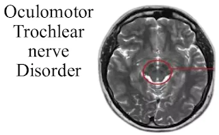
Oculomotor nerve
What is the oculomotor nerve?
The oculomotor nerve is one of the 12 groups of cranial nerves. Large numbers of these nerves are essential for the independent sensory system. The autonomic sensory system gives organs, like your eyes.
- The third cranial nerve is the oculomotor nerve. It allows eye muscle movement, pupil congestion, eye focus and posture of the upper eyelid.
- Cranial nerve III works with other cranial nerves to control eye movements and to support nerve function.
- The olfactory nerve (CN-I) creates a sense of smell.
- Optic nerve (CN II) activates vision.
- The trigeminal nerve (CN-V) empowers the sensation in your face.
- Vestibular and cochlear nerves (CN-VII) create balance and sensitivity.
What is the function of the oculomotor nerve?
It controls four of the six muscles that make eye movements. CN III makes it possible to:
- Lift the upper eyelid.
- Focus on the eyes.
- Respond to light by making the black center of the eye (student) smaller.
- Move your eyes in, out, up and down and control the trauma.
How does CN III work?
Links eye movements to movements that include:
- A place to stay, to focus on something that moves near or far away from you.
- Optokinetic reflex, which shifts the eyes back to their original position after focusing on the object.
- Saccades, a fast movement that changes your look from one object to another.
- Slight pursuit (visual tracking), which enables you to catch your eye on a moving object.
- Vestibule-ocular reflex, which corrects the position of the eyes as your head moves.
- Visual correction, staring at something that does not move.
What is the anatomy of cranial nerve III?
CN III begins in the midbrain. It travels through many structures in your head until it reaches the back of your eyes. Its course includes:
- It comes out in front of the middle brain.
- Passing nearby veins.
- Piercing of thick outer tissue of the brain (dura).
- It enters the cavernous sinus, the empty space behind the nose.
- It leaves a skull in the orbital cavity, a large circular hole behind each eye.
- It connects behind the eye.
- Divided by upper and lower branches.
- The upper and lower limbs are connected by four muscles that control the movement of the eyes, as well as the upper eyelid muscles and the inner muscles that control the student's size and lens focus.
These include:
- Low oblique, which controls eye rolling, upward and outward look.
- Lower rectus, which controls the downward spiral.
- Medial rectus, which controls internal vision.
- Superior rectus, which controls the upward spiral.
- Levator palpebral superiors, which controls your ability to lift your upper eyelid.
- It is the 3rd cranial nerve
- Muscles supplied
- External ocular muscles except lateral rectus and superior oblique
- Levator palpabrae superiors
- Ciliary muscles
- Sphincter pupillae
Etiology (Damage)
- Disseminated sclerosis
- Meningovascular syphilis
- Diabetes mellitus: Pupil will be spared
- Cerebral aneurysms
- Granulomatious disorder: Tolosa Hunt Syndrome
- Oculomotor nerve palsy
Tolosa Hunt Syndrome
Tolosa-Hunt syndrome is a rare disease characterized by periorbital headaches, as well as diminished and painful eye movements (ophthalmoplegia). Symptoms usually affect only one eye (unilateral). In many cases, affected people experience severe acute pain and decreased eye movement.
Symptoms / Signs
- Ptosis
- Diplopia
- External deviation of eye (Divergent strabismus)
- Pupil dilated and not reacting to light or accommodation (complete paralysis)
Trochlear nerve
Course
The trochlear nerve originates in the trochlear nucleus of the brain, which extends from the posterior part of the midbrain (the only cranial nerve exiting the posterior midbrain).
It runs front and down inside the subarachnoid space before piercing the dura mater near the posterior clinoid process of the sphenoid bone.
The nerve travels to the posterior wall of the cavernous sinus (along with the oculomotor nerve, the abducens nerve, the ophthalmic branches, and the maxillary trigeminal nerve and the internal carotid artery) before entering the eye orbit through a high orbital fissure. .
Automotive Function
The trochlear nerve penetrates into a single muscle - the upper oblique, the oculomotor muscle. As the fibers from the trochlear nucleus cross the midbrain before they come out, the trochlear neurones from the contralateral superior oblique are invisible.
The upper oblique tendon is tied to a fibrous structure known as the trochlea, which gives the nerve its name. Although the oblique high-performance procedure is complex, in clinical practice, it is sufficient to understand that the complete action of the upper oblique is to compress and insert the eyeball.
Muscles supplied
Superior oblique muscles
Damage causes
- Strabismus (Squint)
- Paralytic squint: Due to weakness of one or more extra ocular muscles
- Concomitant squint (Lazy eyes): Cause refractive error during childhood
- Nystagmus: Is a series of involuntary, rhythmic oscillation of one or both eyes
- Trochlear nerve palsy
Nystagmus: Series of involuntary, rhythmic oscillations of one or both eyes. It may manifest in horizontal or vertical planes or as a series of rotations about the central axis...
Types:
- Gaze nystagmus
- Fixation nystagmus
- Vestibular nystagmus
- Cerebellar mechanisms


Post a Comment